|
| |||||||||||||||||||||||||||||||||||||||||||||||||||||||||||||||||||
| |||||||||||||||||||||||||||||||||||||||||||||||||||||||||||||||||||
|
Dental Clinic Homepages Dentists Doctors General Lifestyle
Please Fill Our Guest Book |
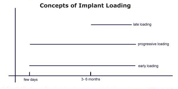
Protocols for immediate or early loading strive for an increase either of
primary stability, which can be achieved either through an optimized implant
form or implant surface or/and optimized surgical preparation of the implant
bed. Conversely, through the modification of the implant surface, an
acceleration of the bony healing is intended to achieve an earlier
osseointegration and therefore a faster acceptable secondary stability for
successful loading. The authors conclude that the use of immediate loading still
consists of a higher risk because of non-existing, not yet defined diagnostic
criteria and can therefore not be recommended to the general practitioner. An
acceptable measurement system may be available in the near future and help
revolutionize the treatment concept in implant therapy.
Osseointegration is the result of a biologic response of the bony tissues to
implants. For oral endosteal implants a three to six month healing period
prior to loading is generally considered a pre-condition for optimal bone
apposition to the fixture. This clinical protocol is based on work done by
Branemark during the 1960's and 1970's when a final proposition for healing
periods for implants of the upper and lower jaw, based on the evaluation of
the surgical and periodontal failure rates, was defined (Br�nemark et al,
1977 [1]
). These protocols on Osseointegration periods were based on early, partially
unfavorable experimental conditions while methods and implant types were
continuously changing:
It is therefore not surprising that in the past, the submerged healing period
was frequently questioned, based on modifications of surgical and prosthetic
procedures. In several animal and human studies the original three to six
month healing period was shortened to an early loading protocol. In other
modifications, implants were immediately loaded on the day of insertion.
Post-Surgical Bone Reaction
Concerning the bone healing after the surgical implant placement, one can draw
the following conclusion:
The perimplant bone reaction is initially the highest after the surgical
trauma and reduces itself slowly over months, returning to the basic turnover.
It is evident that even after the three to six month standard healing protocol
any loading will not be placed on the final bony implant bed but bony
remodeling will still be in progress.
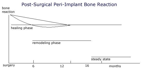
Based on these two conditions it is not feasible to determine a general
healing period depending only on the type of jaw. To define an individual
healing period the bone should be evaluated both during and after the implant
surgery. Because traditional methods are unsuitable for this measurement, new
methods are recently described.
Methods for the evaluation of the peri-implant bone conditions:
Traditional methods Modern methods
The recording of the cutting resistance during the surgery permits some
evaluation of the bony structure (compact versus spongious) along the implant
site. The measurement of the resonance frequency analysis on an inserted
implant provides information about the primary stability between implant and
bone. While primary stability in implantology was previously based on a purely
subjective evaluation, these two new measurement methods permit an objective
recording of the initial implant stability.
Primary and secondary implant stability
To achieve osseointegration an attachment to the bone without any
micro-movement is necessary. Prior dental literature cites micromovement of
the implant as a causative factor in both the formation of an intermediate
layer of connective tissue, which develops between the bone and implant, and
osseointegration failure. Initially, primary stability is entirely mechanical.
During the healing period however, the biologic processes of osseointegration
cause this to change to a mixture of mechanical and biologic stability
(secondary stability).
Protocols for immediate or early loading strive for an increase either of
primary stability, which can be achieved either through an optimized implant
form or implant surface or/and optimized surgical preparation of the implant
bed. Conversely, through the modification of the implant surface an
acceleration of the bony healing is intended to achieve an earlier
osseointegration and therefore a faster acceptable secondary stability (Case
examples Fig. 1 - 12).
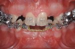
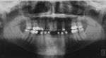
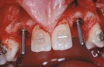
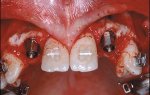
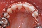
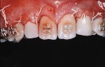
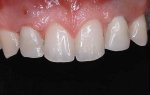
A 19-year-old patient, with multiple genetically missing teeth, at the end of
the preprosthetic orthodontic treatment. In the upper jaw implants are planned
in the area 12 - 25. Implants on 12 and 22 should be immediate loaded because
of esthetic reasons.
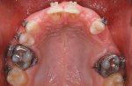
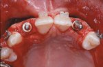
After preparation of the implant bed at the position of both lateral incisors
the prospective implant placement will be marked with a direction indicator.
Two Br�nemark MK IV are inserted and a CeraOne� abutment is definitively
placed on both implants.
Initial implant stability will be measured with the RFA method. The buccal
bony defect will be covered using a membrane technique and a prefabricated
shell-like temporary will be directly, intraorally rebased over the abutment.
Summary
The protocol of successful osseointegration, which was based for over 20 years
on the concept of late loading, is increasingly questioned. In several studies
successful immediate loading or early loading of implants could be shown. Two
indications were described:
The indication for the case selection for immediate loading is based on an
optimal stabilization of the implants through a large number (full, horse shoe
type bridges) or on implants in a very good bone quality or quantity (interforaminal).
Through secondary splinting with the suprastructure, possible micromovement
during the healing period can be prevented in both situations.
All protocols up to the present day however show that the operator based case
selection on subjective criteria. Objective quantitative criteria for case
selection are not yet published. Long term data on immediate, or early implant
loading exist only for interforaminally placed fixtures.
At the moment the use of immediate loading still consists of a higher risk
because of non-existing, not yet defined diagnostic criteria and can therefore
not be recommended to the general practitioner. On the other hand a suitable
measurement system will be available in the near future and might
revolutionize our treatment concept in implant therapy.
References
|
More Articles
Shade selection and Management
Obstructive Sleep Apnea- Do you know about it?
Genetically modified bacteria may prevent cavities - Put you out of Business?
A primer on all composite class materials
Immediate and Early loading of Implants
Placement of gingival restorative margins
Bonding for the New Millennium
Access Cavity Preparation - Molars
New cavity-fighting agent shows promise
Tooth Loss Linked to Pancreatic Cancer in Smokers
2-min brush helps achieve cleaner teeth: Study
Gum disease raises death risk in diabetics: study
Brushing Right After Drinking Soda may Harm Teeth
Benefits of Pre-procedural mouth rinsing?
To Bond Or not to Bond Amalgam
Restoration of endodontic teeth
Options for esthetic restorations
Teeth Whitening Facts and Myths
Fiber reinforced composites in dentistry
Changing concepts in Class I and II cavity preparation
Curing lights for composite resins
Caries Prevention in Children - The Indian Challenge
Mouth Rinsing before dental procedures.
Reviews:
Rotary Endodontic Instrumentation
Direct & Indirect Esthetic Adhesive Restorative Materials
Early and Immediate Loading of Implants
Lasers in Root canal treatment and Endodontics
Abfractions? How they are important in Restorative Dentistry
Latest Research On Dental Pain
| |||||||||||||||||||||||||||||||||||||||||||||||||||||||||||||||||
| |||||||||||||||||||||||||||||||||||||||||||||||||||||||||||||||||||