Preparation of Root Canal
Endoseries 14,
Preparation of Root Canal
Microorganisms are found to a variable degree up to the apical foramen in three modes:
1. as a suspension in the root canal
2. colonizing the canal walls and
3. colonizing the dentinal tubules.
It is impossible to completely sterilize the root canal system due to its complex structure. Luckily for us, removal of the bulk of the microorganisms and their products brings about periradicular healing due to an altered or less pathogenic residual flora.
The aim of canal preparation in vital teeth is to remove pulp tissue, which may become necrotic and infected.
A combined action of mechanical and chemical cleansing is effective in root canal preparation.
Working length determination
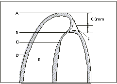
From the preoperative radiograph, estimate the average length of the tooth. Select a reproducible coronal reference point. It should not be part of a portion of tooth or restorative material that is likely to break off. Choose a file that is large enough to be visible on the radiograph (at least size 10).Insert a file into the root canal, 1-2 mm short of the estimated length and take a parallel view radiograph. An average distance of 1 mm short of the radiographic apex is widely accepted as a reasonable estimate of the terminal portion of the canal. Remember, at times, this may be inaccurate by up to 3 mm.(see fig 1)

Relationship between apical foramen, root tip and apical constrication. A=Root apex B= Apical constriction
C= Root canal D=Cementum E=Dentin F=Apical foramen
In cases of narrow root tips, and when there is apical root resorption, working length should be shortened more than 1 mm. In the former, it is because perforation may occur when the root is prepared to a wider diameter(See Fig2) and in the latter, the canal exit may be Ďblunderbussí shaped that can allow extrusion of endodontic materials.

If the tip of the file is short of the radiographic apex by 1 mm, accept it as the working length. If the file is longer than the radiographic apex, measure the distance between the
file tip and a point 1 mm short of the radiographic apex. Subtract this figure from the length of the diagnostic file to get the working length.
If the distance between the file tip and the radiographic apex is greater than 1 mm, subtract 1 mm from this distance and add it to the length of the diagnostic file to get the working length.
If you have reasonable number of endodontic cases, it is worth investing in an electronic apex locator. Many reliable brands are available in the market. I have been using J Moritaís Root ZX to my fullest satisfaction. Endoseries 5 had covered electronic apex locators.
Once you have determined the working length, it is of utmost importance to restrict instrumentation to this length. Avoid displacement of the stops.
The width of the taper to which the canal should be prepared should be based on personal preference and individual clinical experience. If they allow adequate cleaning and obturation, narrowly tapered preparations are more desirable, as they do not compromise root strength and avoid strip perforations.
Mechanical preparation refers to controlled removal of dentin by manipulating root canal instruments. Factors influencing the amount and pattern of dentin removal are,
-design and sharpness of the cutting edge
-the manner in which it is manipulated
-the force applied and
-the operatorís skill.
Operatorís skill is influenced by the ability to discriminate tactile feedback from the instrument and the ability to manipulate the instruments in a controlled way according to the mental image of a three dimensional shape of the root canal system.
You can either rotate the root canal instruments (clockwise and withdraw) or used in a push- pull filing motion to remove dentin. Or you can combine the two (ream and file) by 45-90 degree clockwise movements to engage the dentin and straight pull withdrawal to cut the engaged dentin.
Due to uncontrolled dentin removal, errors in canal preparation viz., ledging, zipping and transportation of apical foramen may result. A reliable method to reduce uncontrolled forces is to use flexible files such as Flex-R, Flexo-file, Nickel-Titanium etc.,
To avoid procedural errors resulting in loss of working length, use smaller instruments (No. 20 or smaller) for sufficient time until the larger sizes pass in the canal without force. You can even create intermediate files as suggested by Weine, by trimming 1 m from the tip of the file and rounding off sharp edges on a diamond nail file. This way you can convert #10, 15& 20 files to # 12,17 and 22
Ref:
1. Christopher J R Stock, Gulabivala K, Walker T, Goodman JR: Endodontics, 2nd Ed. NewYork, Mosby- Wolfe p 97-144, 1995.
2. Weine FS: Endodontic Therapy 3rd Ed. Mosby, 1982